Skip to Dynamap
Team
Publications
News
Research Summary
DYNAMAP
(Dynamical Brain Mapping and Pathophysiology of Focal Human Epilepsies)
The main research themes of the DynaMaP team are i) the methodology of brain mapping for epilepsy and cognition (based on EEG, magnetoencephalography and intracerebral EEG) ii) the pathophysiology of human epilepsy. A key feature of the team is to be composed of people with diverse backgrounds (engineers, clinicians, neuroscientists) and to be located within the Timone hospital, which ensures tight collaboration between research and clinics. In the past years, we notably obtained results on the characterization of epileptic networks and on the simultaneous recording of MEG an intracerebral EEG.
We are part of large-scale projects (RHU Epinov, ERC Galvani). Moreover, the Dynamap team runs the magnetoencephalography platform, which has the IBISA and “AMU platform” labels, is member of France Life Imaging network, and of several AMU Institutes (NeuroMarseille, Laënnec, Marseille Imaging).
A particular effort is made towards translation of our work to clinicians and researchers through the multi-platform Anywave software and its associated plugins (epileptogenicity Index, Delphos, Gardel…), which architecture specifically aims at allowing fast implementation of algorithms to the end users.
We obtained recently an A*midex industrial chair, “NewMeg Marseille”, in collaboration with the Meg4Health company.
DYNAMAP TEAM
TEAM LEAD
Christian BÉNAR
DR, INSERM
EMAIL: christian.benar@univ-amu.fr
PHONE: +33 4 91 38 55 77
I graduated from Ecole Supérieur d'Electricité (Supélec) in 1994. I then spent one year as an engineer at the Hospital Saint-Anne in Toulon (with Franck Vidal), and two years as a programmer at Stellate Systems (Montréal, Dir Jean Gotman). I did my PhD under the supervision of Jean Gotman at the Montreal Neurological Institute (MNI). Back to France in 2004, my postdocs were in Marseille (fMRI Center, with Jean-Luc Anton) and in Sophia Antipolis (Maureen Clerc and Theodore Papadopoulo).
I was appointed researcher Inserm (CR1) in 2006, and Inserm Director of Research (DR2) in 2018 . Since January 2012, I am the leader of the "Dynamical Brain Mapping Group” here at INS. Since September 2014, I am scientific head of the Marseille MEG platform. Since 2023, I am a member of the scientific council of the Fédération Française de Recherche sur l’épilepsie (FFRE) and elected member of the Neurotechnology Task force of international League against Epilepsy (ILAE).
My research interest is signal processing applied to brain signals (EEG, MEG, intracerebral EEG), in order to characterize the spatio-temporal dynamics of networks in cognition and disease. For more information, see my CV .
TEAM MEMBERS
DYNAMAP PUBLICATIONS
Selected Publications
Jedynak, M., et al. (2023) “Variability of Single Pulse Electrical Stimulation Responses Recorded with Intracranial Electroencephalography in Epileptic Patients“ Brain Topography
López‐Madrona, Víctor J., et al. "Magnetoencephalography can reveal deep brain network activities linked to memory processes." Human Brain Mapping 43.15 (2022): 4733-4749.
Simula, S., Daoud, M., Bénar, C. G., Benquet, P., Ruffini, G., Wendling, F., & Bartolomei, F. (2022). Transcranial current stimulation in epilepsy: A systematic review of the fundamental and clinical aspects. Frontiers in Neuroscience, 1289.
Bénar, C. G., Velmurugan, J., López-Madrona, V. J., Pizzo, F., & Badier, J. M. (2021). Detection and localization of deep sources in magnetoencephalography: A review. Current Opinion in Biomedical Engineering, 18, 100285.
Pizzo F, Roehri N, Medina Villalon S, Trébuchon A, Chen S, Lagarde S, Carron R, Gavaret M, Giusiano B, McGonigal A, Bartolomei F, Badier JM, Bénar CG. Deep brain activities can be detected with magnetoencephalography. Nat Commun. 2019 Feb 27;10(1):971.
Lagarde S, Roehri N, Lambert I, Trebuchon A, McGonigal A, Carron R, Scavarda D, Milh M, Pizzo F, Colombet B, Giusiano B, Medina Villalon S, Guye M, Bénar CG, Bartolomei F. Interictal stereotactic-EEG functional connectivity in refractory focal epilepsies. Brain. 2018 Oct 1;141(10):2966-2980.
Roehri N, Pizzo F, Lagarde S, Lambert I, Nica A, McGonigal A, Giusiano B, Bartolomei F, Bénar CG. High-frequency oscillations are not better biomarkers of epileptogenic tissues than spikes. Ann Neurol. 2018 Jan;83(1):84-97.
Medina Villalon S, Paz R, Roehri N, Lagarde S, Pizzo F, Colombet B, Bartolomei F, Carron R, Bénar CG. EpiTools, A software suite for presurgical brain mapping in epilepsy: Intracerebral EEG. J Neurosci Methods. 2018 Jun 1;303:7-15.
Bartolomei F, Lagarde S, Wendling F, McGonigal A, Jirsa V, Guye M, Bénar C. Defining epileptogenic networks: Contribution of SEEG and signal analysis. Epilepsia. 2017 Jul;58(7):1131-1147. doi: 10.1111/epi.13791. Epub 2017 May 20. Review.
Latest Publications

DYNAMAP NEWS
DYNAMAP RESEARCH
Simultaneous recordings of MEG, EEG and SEEG
Most previous work on the comparison of MEG, EEG and intracerebral EEG was performed on separate recordings. However, this is far from being optimal. Indeed, only simultaneous recordings of surface and depth activity permit to investigate the exact same activity, independently of spontaneous fluctuations that can occur from a session to another (depending on subject state, medication etc). Moreover, simultaneous recordings allow using interevent fluctuations as a source of information on the link between signals, as was done on simultaneous EEG-fMRI.
We have shown for the first time the feasibility of trimodal simultaneous recordings: EEG, MEG and SEEG (Dubarry et al., 2014). Technical developments have been proposed in (Badier et al., 2017) and an application to presurgical mapping in (Gavaret et al., 2016). Recently, thanks to the simultaneous intracerebral recordings, we have shown that it is possible to capture the activity of deep structures on MEG (Pizzo et al 2019, López-Madrona et al. 2022).
Activity evoked by visual stimulation (checkerboard) on MEG, EEG and intracerebral EEG (SEEG) recorded simultaneously (Dubarry et al 2014).
Visibility of deep mesial source on an ICA component of MEG (Pizzo et al 2019).
Pizzo F, Roehri N, Medina Villalon S, Trébuchon A, Chen S, Lagarde S, Carron R, Gavaret M, Giusiano B, McGonigal A, Bartolomei F, Badier JM, Bénar CG. Deep brain activities can be detected with magnetoencephalography. Nat Commun. 2019 Feb 27;10(1):971.
Brain networks in epilepsy and sleep
An active line of research of the team, incollaborations between clinicians and researches, aims at characterizing brain networks from both intracerebral EEG and from surface (MEG, EEG) signals. The network view enables to better understand mechanisms of epilepsy and sleep; it also provides new venues for defining markers of the epileptogenic zone. Methods are available in the Anywave software, Epitools and other plugins (see below “Anywave” section).
Lambert I, Tramoni-Negre E, Lagarde S, Roehri N, Giusiano B, Trebuchon-Da Fonseca A, Carron R, Benar CG, Felician O, Bartolomei F.
Hippocampal Interictal Spikes during Sleep Impact Long-Term Memory Consolidation. Ann Neurol. 2020 Jun;87(6):976-987.
McGonigal A, Marquis P, Medina S, Bartolomei F, Rheims S, Bernard C, Bénar C. Postictal stereo-EEG changes following bilateral tonic-clonic seizures. Epilepsia. 2019 Aug;60(8):1743-1745.
Bartolomei F, Lagarde S, Scavarda D, Carron R, Bénar CG, Picard F. The role of the dorsal anterior insula in ecstatic sensation revealed by direct electrical brain stimulation. Brain Stimul. 2019 Sep-Oct;12(5):1121-1126.
Lagarde S, Roehri N, Lambert I, Trebuchon A, McGonigal A, Carron R, Scavarda D, Milh M, Pizzo F, Colombet B, Giusiano B, Medina Villalon S, Guye M, Bénar CG, Bartolomei F. Interictal stereotactic-EEG functional connectivity in refractory focal epilepsies. Brain. 2018 Oct 1;141(10):2966-2980.
Lambert I, Roehri N, Giusiano B, Carron R, Wendling F, Benar C, Bartolomei F. Brain regions and epileptogenicity influence epileptic interictal spike production and propagation during NREM sleep in comparison with wakefulness. Epilepsia. 2018 Jan;59(1):235-243
Jmail N, Gavaret M, Bartolomei F, Chauvel P, Badier JM, Bénar CG. Comparison of Brain Networks During Interictal Oscillations and Spikes on Magnetoencephalography and Intracerebral EEG. Brain Topogr. 2016 Sep;29(5):752-65.
Courtens S, Colombet B, Trébuchon A, Brovelli A, Bartolomei F, Bénar CG. Graph Measures of Node Strength for Characterizing Preictal Synchrony in Partial Epilepsy. Brain Connect. 2016 Sep;6(7):530-9.
Bartolomei F, Trébuchon A, Bonini F, Lambert I, Gavaret M, Woodman M, Giusiano B, Wendling F, Bénar C. What is the concordance between the seizure onset zone and the irritative zone? A SEEG quantified study. Clin Neurophysiol. 2016 Feb;127(2):1157-1162.
Time frequency methods for high-frequency activity
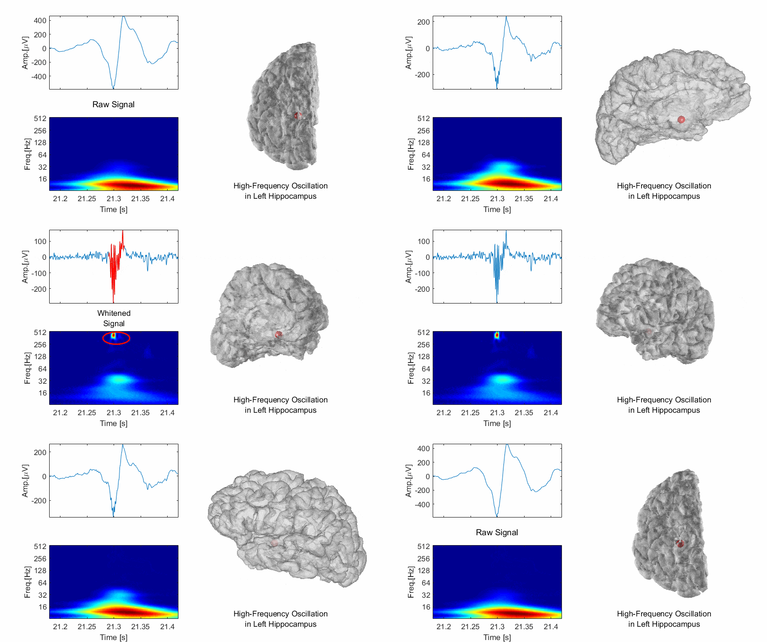
High frequency oscillations (HFOs) have been proposed as a new marker for delineating epileptic tissues. One difficulty in characterizing these activity is that they are of much smaller amplitude than classical spike markers. We have proposed a new method to whiten the SEEG signals in order to better visualize and detect HFOs (Roehri et al., 2016). In a latter publication, we have shown that it may be useful to combine spike and HFO information instead of considering them separately (Roehri et al., 2018). The Delphos plugin is available here.
Effect of whitening on SEEG traces (see Roehri et al 2016).
Roehri N, Lina JM, Mosher JC, Bartolomei F, Benar CG. Time-Frequency Strategies for Increasing High-Frequency Oscillation Detectability in Intracerebral EEG. IEEE Trans Biomed Eng. 2016 Dec;63(12):2595-2606.
Roehri N, Pizzo F, Bartolomei F, Wendling F, Bénar CG. What are the assets and weaknesses of HFO detectors? A benchmark framework based on realistic simulations. PLoS One. 2017 Apr 13;12(4):e0174702.
Roehri N, Pizzo F, Lagarde S, Lambert I, Nica A, McGonigal A, Giusiano B, Bartolomei F, Bénar CG. High-frequency oscillations are not better biomarkers of epileptogenic tissues than spikes. Ann Neurol. 2018 Jan;83(1):84-97.
Scholly J, Pizzo F, Timofeev A, Valenti-Hirsch MP, Ollivier I, Proust F, Roehri N, Bénar CG, Hirsch E, Bartolomei F. High-frequency oscillations and spikes running down after SEEG-guided thermocoagulations in the epileptogenic network of periventricular nodular heterotopia. Epilepsy Res. 2019 Feb;150:27-31
Jmail N, Gavaret M, Bartolomei F, Bénar CG. Despiking SEEG signals reveals dynamics of gamma band preictal activity. Physiol Meas. 2017 Feb;38(2):N42-N56.
The AnyWave software and plugins
Within the DynaMap team, we have developed the AnyWave software for visualizing electrophysiological traces (main developer Bruno Colombet). This software is multiplatform and modular, and in particular, permits easy addition of plugins in Python or Matlab (Colombet et al., 2015). In addition, our software Gardel permits to register and segment SEEG electrodes (Medina Villalon et al., 2018). The AnyWave software is available for download here and the plugins there.
Example of ICA decomposition in Anywave (MEG traces). Software available on MEG wiki.
Colombet B, Woodman M, Badier JM, Bénar CG. AnyWave: a cross-platform and modular software for visualizing and processing electrophysiological signals. J Neurosci Methods. 2015 Mar 15;242:118-26.
Medina Villalon S, Paz R, Roehri N, Lagarde S, Pizzo F, Colombet B, Bartolomei F, Carron R, Bénar CG. EpiTools, A software suite for presurgical brain mapping in epilepsy: Intracerebral EEG. J Neurosci Methods. 2018 Jun 1;303:7-15.
